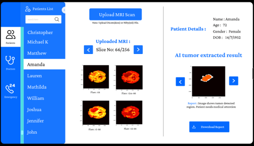
We trained a 3D convolutional neural network on a dataset of thousands of annotated MRI scans, achieving over 94% sensitivity for gliomas, meningiomas, and pituitary tumors. The model processes a full 3D MRI scan in under 90 seconds and outputs a detailed heatmap overlaid on the original scan, showing the precise location, estimated size, and shape of any detected anomalies.
3D MRI Scan Submission
The platform accepts DICOM-format 3D MRI files via a secure, HIPAA-compliant upload portal. Scans are preprocessed automatically - skull-stripped, registered to MNI space, and normalized - before being passed to the detection model. The entire preprocessing pipeline runs in the cloud, requiring no local compute resources from the clinical team.
AI Tumor Detection
A 3D U-Net architecture performs voxel-level segmentation of the MRI volume, identifying regions that match the morphological signatures of known tumor types. The model was trained on diverse, multi-institution datasets to ensure robustness across different MRI scanner manufacturers and acquisition protocols.
Detailed Tumor Report
The AI generates a structured clinical report containing the tumor's probable type and grade, estimated volume in cubic centimeters, and a 3D bounding outline overlaid on the original scan for radiologist review. Findings are presented on an interactive web viewer built on the Cornerstone.js DICOM viewer framework.
Enhanced Medical Support
NeuroScan AI is designed as a decision-support tool, not a replacement for clinical judgment. All AI findings are presented alongside confidence scores and flagged for radiologist confirmation before any clinical action is taken. An integrated feedback loop allows radiologists to correct predictions, continuously improving model performance over time.